Robert Johnston spent months at a microscope watching something that, by the textbook he trained on, was not supposed to happen.
The tissue in question was the foveola, the pinhead at the dead center of the retina that does a wildly outsized share of the work. It is a speck, but the Johns Hopkins team estimates it delivers roughly 50 percent of what your brain registers as sight; the sharp central vision you are using to read this sits there and almost nowhere else. What makes it sharp is what it lacks. The foveola is packed with red and green cones, the photoreceptors that carry fine detail and most of color, and the blue cones that blanket the rest of the retina are, in that one spot, gone.
For about thirty years the field had a tidy explanation for their absence. The blue cones appear early, the story went, the way they do everywhere else, then drift out of the way to the periphery and stay blue cones forever. “The main model in the field from about 30 years ago was that somehow the few blue cones you get in that region just move out of the way,” Johnston told reporters. It was a reasonable story. It was also, his lab now reports in the Proceedings of the National Academy of Sciences, the wrong one.
The blue cones did not migrate. They became something else.
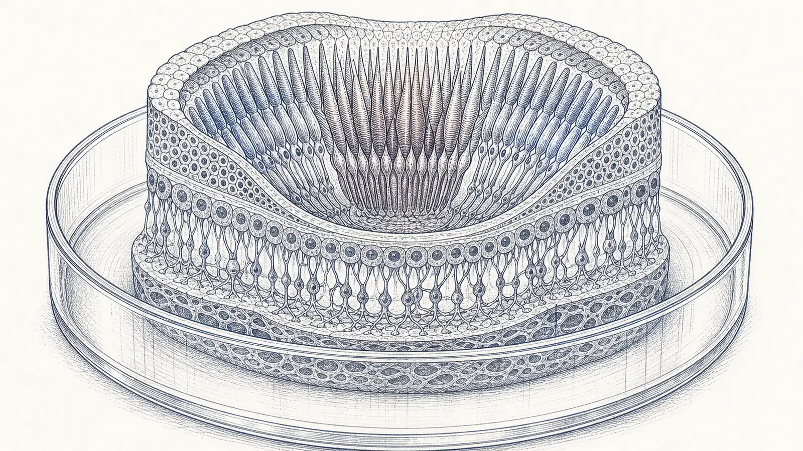
Johnston’s group reached that conclusion by growing human retinas from scratch. Using stem cells, they cultured retinal organoids, self-organizing clusters of lab-grown tissue that mimic the developing eye, and watched them over the months it takes a fovea to pattern itself. Around weeks 10 to 12 of the equivalent fetal window, a sparse population of blue cones appeared in the foveolar region, exactly as expected. By week 14, on the timeline the organoids reproduced, those cells were gone, and red and green cones stood where they had been. Nothing wandered off. The cells changed identity in place.
Two signals did the work, in sequence. First, retinoic acid, a molecule the body makes from vitamin A, gets broken down in the foveola, and that local scarcity chokes off the formation of new blue cones. Then thyroid hormone moves in and pushes the blue cones that remain to convert into red and green ones. “First, retinoic acid helps set the pattern,” Johnston said. “Then, thyroid hormone plays a role in converting the leftover cells. That’s very important, because if you have those blue cones in there, you don’t see as well.”
The thyroid half of that mechanism did not come out of nowhere. Johnston’s lab has been circling it for years. In 2018, working in the same organoid system, the group reported in Science that thyroid hormone signaling is the switch that decides cone identity across the whole retina: low signaling early specifies blue cones, high signaling late produces red and green ones, with the retina running its own enzymes to degrade the hormone at one stage and activate it at another. The new paper takes that switch and shows it operating in the one place where the stakes for human vision are highest, alongside a retinoic-acid step the earlier work had not pinned down. It is the rare case of a lab confirming its own hunch rather than a competitor’s.
If you only read the headlines, you might come away thinking the lesson is to buy vitamin A. The study traveled into the news under the word “vitamin,” and retinoic acid is a vitamin A derivative, so the phrasing is not technically wrong. It is just an invitation to the wrong conclusion. Nothing here suggests that swallowing more vitamin A sharpens a grown eye or rescues a failing one. The retinoic acid in this story is a signaling molecule inside a fetal retina, dialed up and down at a particular place in a particular week, not something you take with breakfast. The finding is about how the eye builds itself, not about anything on a pharmacy shelf.
What the work does open, at least in principle, is the door the field has been leaning on for a decade: making photoreceptors to order. If you know the exact molecular choreography that turns a cell into a red or green cone, you can imagine directing stem cells to produce them on demand and transplanting them into retinas damaged by macular degeneration and other age-related disease. “The goal would be to eventually make an almost made-to-order population of photoreceptors” for cell-replacement therapy, said Katarzyna Hussey, the study’s first author. That is the ambition, and it is a serious one. It is also cells in a dish. An organoid is a remarkable stand-in for a developing eye and still not an eye, and no patient’s vision has been restored by any of this. The distance between a correctly patterned foveola in a culture plate and a working transplant in a human retina is measured in years, and in trials that have not been run.
None of which touches the smaller, cleaner result sitting underneath the therapy talk. A claim that a lot of people believed for thirty years, printed in a lot of textbooks, describes a migration that never happens. The cells never went anywhere. Johnston just grew the tissue slowly enough to watch them turn into something else where they sat.
Sources
- PNAS – Hussey, Johnston et al., retinoic acid and thyroid hormone pattern human foveolar cones (2026; 123(7))
- Johns Hopkins Hub – Lab-grown retinas show how humans develop sharp vision
- Neuroscience News – How the eye develops sharp vision (quotes; foveola’s share of sight)
- ScienceDaily – A vitamin A discovery is changing what scientists know about vision
- Science – Eldred et al., thyroid hormone signaling specifies cone subtypes in human retinal organoids (2018)
- SciTechDaily – New research challenges 30-year-old theory of eye development (funding sources)